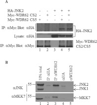
Figure 1. WDR62 association with JNK2 and MKK7.
(A) WDR62 CS2 and CS5 splice variants associate with JNK2. HEK-293T cells were transfected with Myc-WDR62 CS2, Myc-WDR62 CS5 and HA–JNK2 in various combinations as indicated. Cell lysates were subjected to immunoprecipitation (IP) with anti-Myc antibodies followed by Western blotting with either anti-HA or anti-Myc antibodies (top panel and bottom panel respectively). The expression level of HA–JNK2 was determined by blotting total cell lysate with anti-HA antibody (middle panel). (B) Co-immunoprecipitation of endogenous WDR62 with JNK and MKK7. Endogenous WDR62 was immunoprecipitated from HEK-293T cells using anti-WDR62 3G8 antibodies. Monoclonal anti-HA antibodies were used as a negative control (lane 2). Eluted proteins were analysed by Western blotting with either anti-JNK (upper panel) or anti-MKK7 (lower panel) antibodies. Cell lysate (10% of total) was used to assess the expression level of JNK1/2 and MKK7 in the cells (lane 1). Anti-(rabbit light chain) antibody linked to HRP was used as a secondary antibody. Protein molecular mass markers (in kDa) are indicated on the left-hand side. The anti-HA and anti-WDR62 monoclonal antibodies were loaded directly into lanes 4 and 5 respectively to demonstrate lack of cross-reactivity of the anti-rabbit secondary antibody with the mouse IgG heavy chain.

