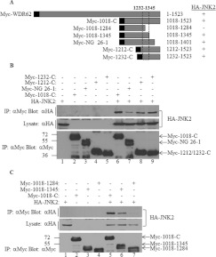
Figure 3. JNK association is mapped to a 113-amino-acid region within WDR62 at position 1232–1345.
(A) Schematic representation of the WDR62 deletion constructs used in the experiments. The black square represents the position of the Myc epitope tag. Summary of the binding of the various WDR62 fragments to HA–JNK2 is indicated by + and −. The broken box indicates the minimal region that preserves JNK binding. (B and C) HEK-293T cells were transfected with a series of plasmids encoding various Myc-tagged WDR62 C-terminal fragments together with HA–JNK2 as indicated. Cell lysates were subjected to immunoprecipitation (IP) with anti-Myc antibodies followed by Western blotting with either anti-HA or anti-Myc antibodies (top panel and bottom panel respectively). Myc-1018-C was used as a positive control for the HA–JNK2 interaction. The expression level of HA–JNK2 was determined by blotting the total cell lysate with anti-HA antibody (middle panel). The migration of the relevant proteins is indicated by arrows. Molecular mass markers (in kDa) are indicated on the left-hand side.

