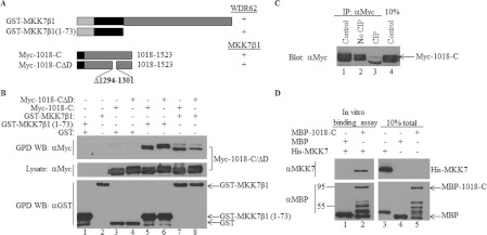
Figure 8. MKK7β1 N-terminal extension associates directly with WDR62 independently of JNK.
(A) Schematic representation of the GST-fused MKK7β1 constructs and Myc-1018-C constructs used in the experiments. The black rectangle represents the MKK7β1 N-terminal spliced fragment encoding amino acids 1–73. The black square represents the position of the Myc epitope tag. The binding of the various WDR62 fragments to MKK7 is indicated by +/−. (B) HEK-293T cells were transfected with plasmids encoding Myc-tagged wild-type or ΔD mutant WDR62 fragments together with the full-length or N-terminal fragment of MKK7β1 fused to GST as indicated. An empty GST plasmid was used as a negative control. GST-containing complexes were isolated from cell lysates with glutathione–agarose beads, washed extensively and eluted with reduced glutathione. The protein complexes were subjected to Western blotting (WB) with either anti-Myc or anti-GST antibodies (upper and lower panels respectively). The expression level of the WDR62 fragments was determined by blotting the total cell lysate with anti-Myc antibody (middle panel). The migration of the relevant proteins is indicated by arrows. (C) HEK-293T cells were transfected with the Myc-1018-C expression plasmid. The WDR62 fragment was immunoprecipitated (IP) from the cell lysate with anti-Myc antibody and either eluted directly in sample buffer (lane 1) or resuspended in CIP buffer in the absence (lane 2) or presence (lane 3) of CIP. The samples were incubated at 37°C for 30 min. Eluted proteins were subjected to SDS/PAGE and Western blotting with anti-Myc antibody. The expression level of the WDR62 fragment was determined by blotting the total cell lysate with anti-Myc antibody (lane 4). (D) Bacterially purified MBP, MBP-1018-C and His–MKK7 proteins were mixed as indicated for 2 h at 37°C. Subsequently, proteins were attached to amylose resin, washed extensively and then eluted using maltose. The protein complexes were subjected to Western blot analysis with either anti-MKK7 (upper panel) or anti-MBP (lower panel) antibodies. The migration of the relevant proteins is indicated by arrows. Molecular mass markers (in kDa) are indicated on the left-hand side. GPD, GST pull-down.

