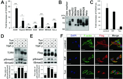
Figure 1. N-linked glycosylation levels of TβRII regulate TGF-β signalling activity and subcellular localization of TβRII.
(A) Cells were transfected with the pGL3-(CAGA)12-luciferase reporter gene and pCMV-βgal. At 12 h after transfection, TGF-β1 (5 ng/ml) was added to the medium for 16 h. Cells were then collected for luciferase (luc.) and β-galatosidase assays. Results represent luciferase activity relative to β-galactosidase activity and are means±S.D. for experiments in triplicate. ***P<0.001; *P<0.05; ns, not significant. (B) FLAG–TβRII was transfected into various cell lines. After 30 h of transfection, cells were harvested and immunoblotted with an anti-FLAG antibody. (C) (CAGA)12 and β-galatosidase were transfected into the A549 cell line. At 6 h after transfection, A549 was treated with KIF (10 μg/ml for 24 h) or Tun (1 μg/ml for 12 h), followed by incubation with or without TGF-β1 (5 ng/ml for 16 h). Cells were then collected for luciferase and β-galatosidase assays. Results are means±S.D. for experiments in triplicate. Note that inhibiting N-linked glycosylation of TβRII by KIF or Tun treatment led to decreased (CAGA)12-luciferase transcriptional activity. (D and E) Untransfected (D) or FLAG–TβRII-transfected (E) A549 was treated with KIF or Tun, followed by TGF-β1 treatment (5 ng/ml for 30 min). Cell extracts were immunoblotted with anti-FLAG, anti-phospho-Smad2 or anti-Smad2 antibody. Band intensities representing phospho-Smad2 and Smad2 expression levels were converted into densitometry using ImageJ software in the ratio of phospho-Smad2 to Smad2. Note that KIF or Tun treatment reduced or inhibited the N-linked glycosylation level of TβRII as well as Smad2 phosphorylation. (F) Fluorescence micrographs of HeLa cells that were transiently transfected with FLAG-tagged TβRII and untreated or treated with KIF or Tun. Cells were stained with an anti-FLAG antibody (red) and phalloidin (F-actin; green). Note that TβRII proteins are mainly localized on the cell surface in the untreated and KIF-treated HeLa cells, whereas they accumulated mostly in the perinuclear region in the Tun-treated cells. DAPI was used for nuclear staining (blue). Scale bars, 50 μm.

