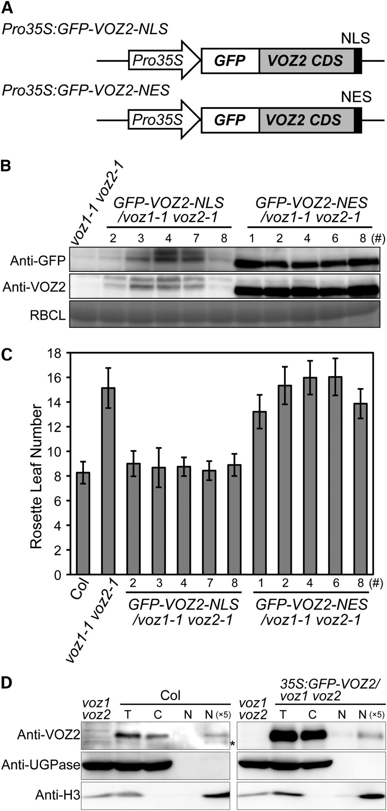
Figure 5.
Subcellular Localization Analysis of Functional VOZ2.
(A) Diagrams of GFP-VOZ2 constructs with NLS or NES.
(B) GFP-VOZ2 protein levels in the seedlings on day 10. Total soluble proteins were subjected to protein immunoblot analysis with anti-GFP and anti-VOZ2 antibodies. Coomassie blue staining of ribulose-1,5-bisphosphate carboxylase/oxygenase large subunit (RBCL) is shown as a loading control.
(C) Rosette leaf number at bolting of NLS and NES lines grown under LD conditions. Data are the mean ± sd (n ≥ 27).
(D) Protein gel blot of cytosolic and nuclear fractions of Col and the Pro35S:GFP-VOZ2/voz1 voz2 line grown under continuous white light for 10 d was probed with anti-VOZ2, anti-UGPase, and anti-histone H3 antibodies. Asterisk represents nonspecific detection. C, cytosolic fraction; N, nuclear fraction; N (×5), fivefold concentrated nuclear fraction; T, total fraction.

