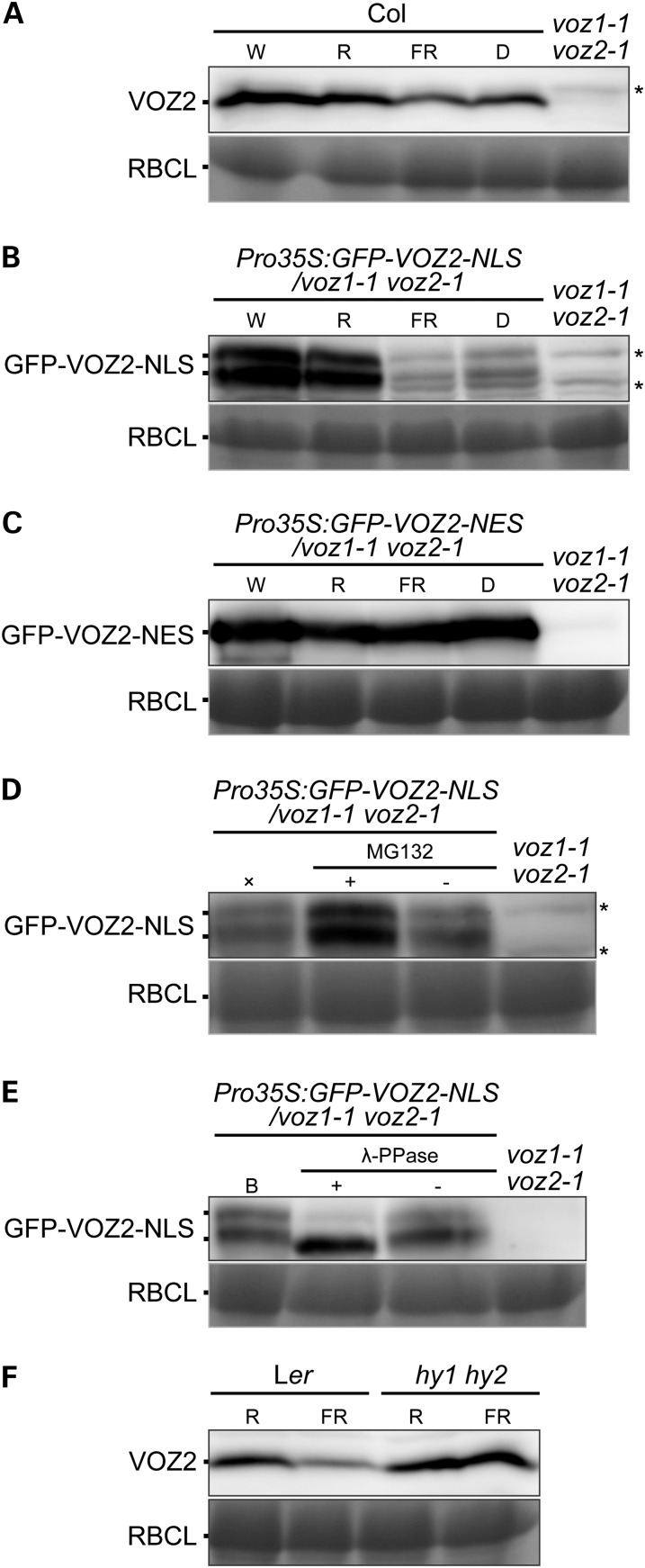
Figure 7.
Degradation and Phosphorylation of VOZ2 Protein.
Protein immunoblotting with anti-VOZ2 antibodies. Coomassie blue staining of ribulose-1,5-bisphosphate carboxylase/oxygenase large subunit (RBCL) is shown as a loading control. Asterisks represent nonspecific detection.
(A) to (C) Protein levels of VOZ2, GFP-VOZ2-NLS, and GFP-VOZ2-NES in Col (A), Pro35S:GFP-VOZ2-NLS/voz1 voz2 line #7 (B), and Pro35S:GFP-VOZ2-NES/voz1 voz2 line #8 (C), respectively. Plants were grown under continuous white light for 10 d and treated with either white (W), red (R), far-red (FR) light, or darkness (D) for 24 h. Each lane contained 60 μg (A), 100 μg (B), or 50 μg (C) of total proteins.
(D) Seedlings grown under continuous white light for 10 d were pretreated with (+) or without (−) 50 μM MG132 for 3 h and transferred to far-red light for 12 h. ×, a control treated with only far-red light. Each lane contained 100 μg of total proteins.
(E) Proteins were extracted from 10-d-old seedlings under continuous white light and incubated with (+) or without (−) λ-PPase. A control sample before λ-PPase treatment is indicated by the letter B. Each lane contained 100 μg of total proteins.
(F) VOZ2 protein levels in Ler and hy1 hy2 mutant. Plants were grown under continuous white light for 10 d and treated with either red or far-red light for 24 h. Each lane contained 65 μg of total proteins.

