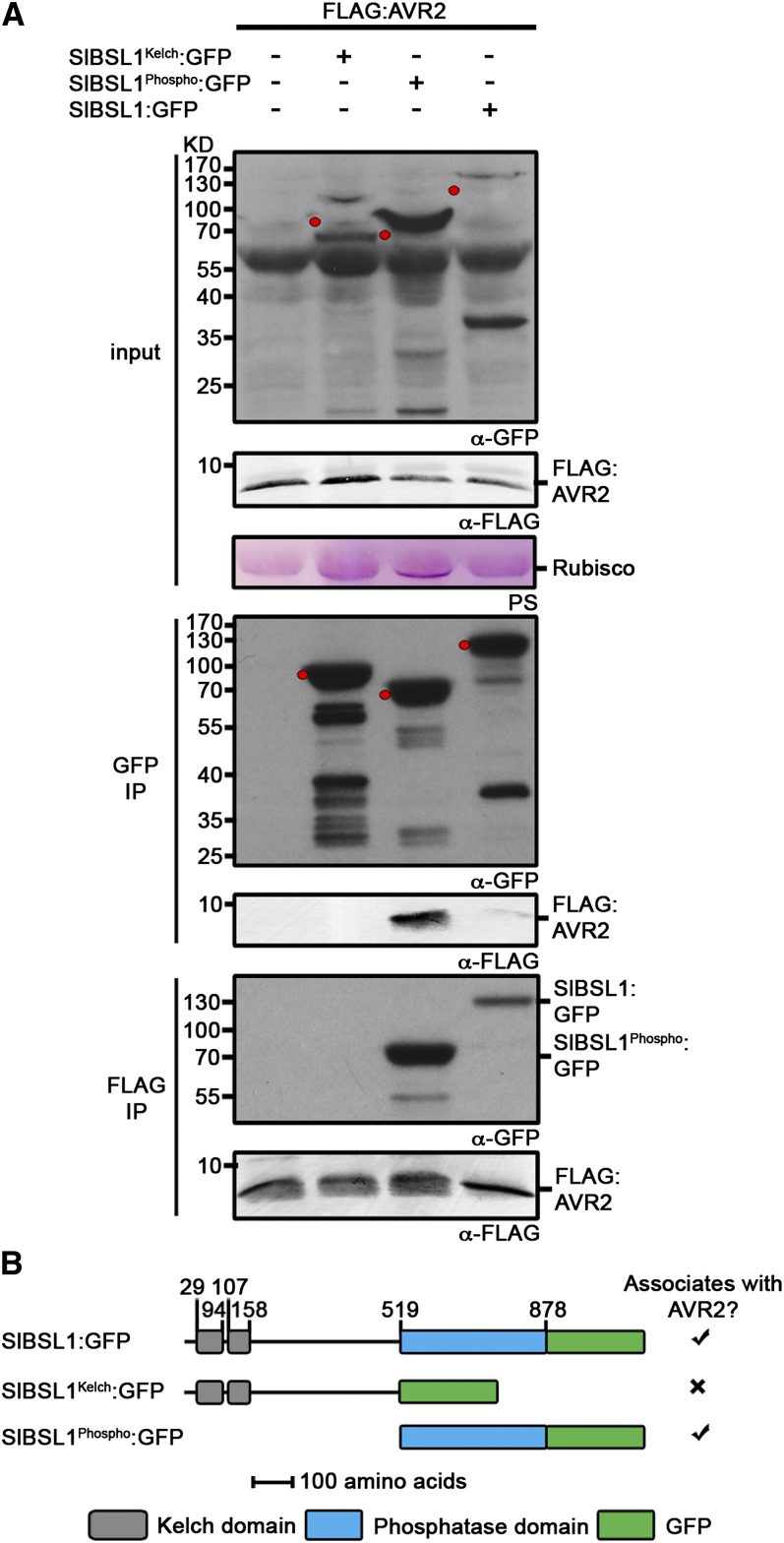
Figure 2.
Immunoblots Showing AVR2 Specifically Associates with the Putative Phosphatase Domain of Sl-BSL1 in Planta.
(A) FLAG:AVR2 was transiently expressed alone, with SlBSL1Kelch:GFP, SlBSL1Phospho:GFP, or SlBSL1:GFP in N. benthamiana. Immunoprecipitates (IP) obtained with anti-FLAG or anti-GFP antiserum and total protein extracts were immunoblotted with appropriate antisera. The expected sizes of the GFP fusion proteins are indicated by red dots in the crude extracts and GFP co-IP probed with anti-GFP antibody. PS, Ponceau stain; Rubisco, ribulose-1,5-bisphosphate carboxylase/oxygenase.
(B) Schematic illustrating the regions of Sl-BSL1 used to assess interaction with AVR2. Numbers indicate position of the amino acid residues.

