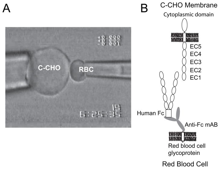
FIGURE 1.
A, configuration of the CHO cells and modified red blood cell in the micropipette measurements. The CHO cell (left) is aspirated into a 7-μm diameter glass pipette. The CHO expresses the full-length cadherin, including the cytoplasmic, transmembrane, and extracellular regions. The red blood cell (right) is aspirated into a 1.3-μm diameter pipette. The RBC, which is activated with CrCl3 binds capture antibody to the cell surface. The capture antibodies used were either monoclonal anti-His6 or monoclonal anti-human-Fc antibodies. These in turn capture the hexahistidine-tagged or Fc-tagged soluble cadherin ectodomains. B, scheme illustrating the relative protein configurations on the opposing cells. The full-length C-cadherin on the C-CHO (top) faces the recombinant cadherin ectodomain bound to the RBC surface (bottom). This illustrates the CEC1–5 Fc captured by the monoclonal anti-human Fc antibody, which is covalently bound to a glycoprotein on the RBC.

