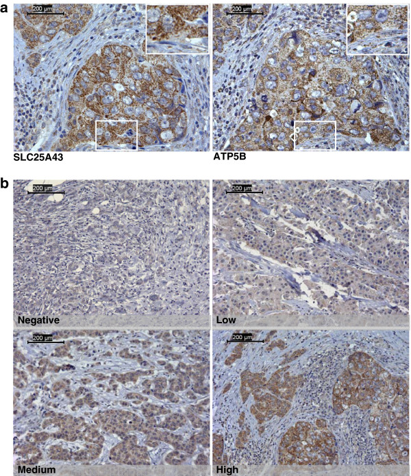
Figure 1.
Protein expression of SLC24A43 in HER2-positive tumours on TMA. Staining of SLC25A43 is localised to the cytoplasm of the cells. Comparison of staining localisation and pattern between SLC25A43 and the mitochondrial inner membrane protein ATP5B in breast tumours (x40 Objective) (a). The four different levels of SLC24A43 expression in HER2-positive tumours used for scoring (x20 Objective) (b). Scale bar is showing 200 μm.

