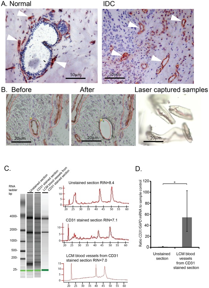
Figure 2. Combined CD31-immunostaining and LCM of blood vessels in breast tissue.
(A) Normal and IDC breast sections were stained with anti-CD31 monoclonal antibody using our modified staining protocol. Blood vessels are stained in red with CD31-AEC (Arrowheads, blood vessels), and cell nuclei are stained in blue with Cresyl Violet. (B) PALM laser microdissection of CD31-positive blood vessels. (C) Agilent bioanalyser of RNA from LCM blood vessels compared with unstained control and CD31 stained sections. RNA quality was rated according to the RNA integrity number (RIN). Histograms correspond to RNA bioanalyser profiles. (D) Real-time PCR of CD31 mRNA expression in LCM blood vessels from breast tissue confirms high level of CD31 in laser captured material. CD31 mRNA expression was given as a ratio to G6PD mRNA (internal control), and the data represented relative to unstained sample control (n = 2, ± fold range, *p<0.05).

