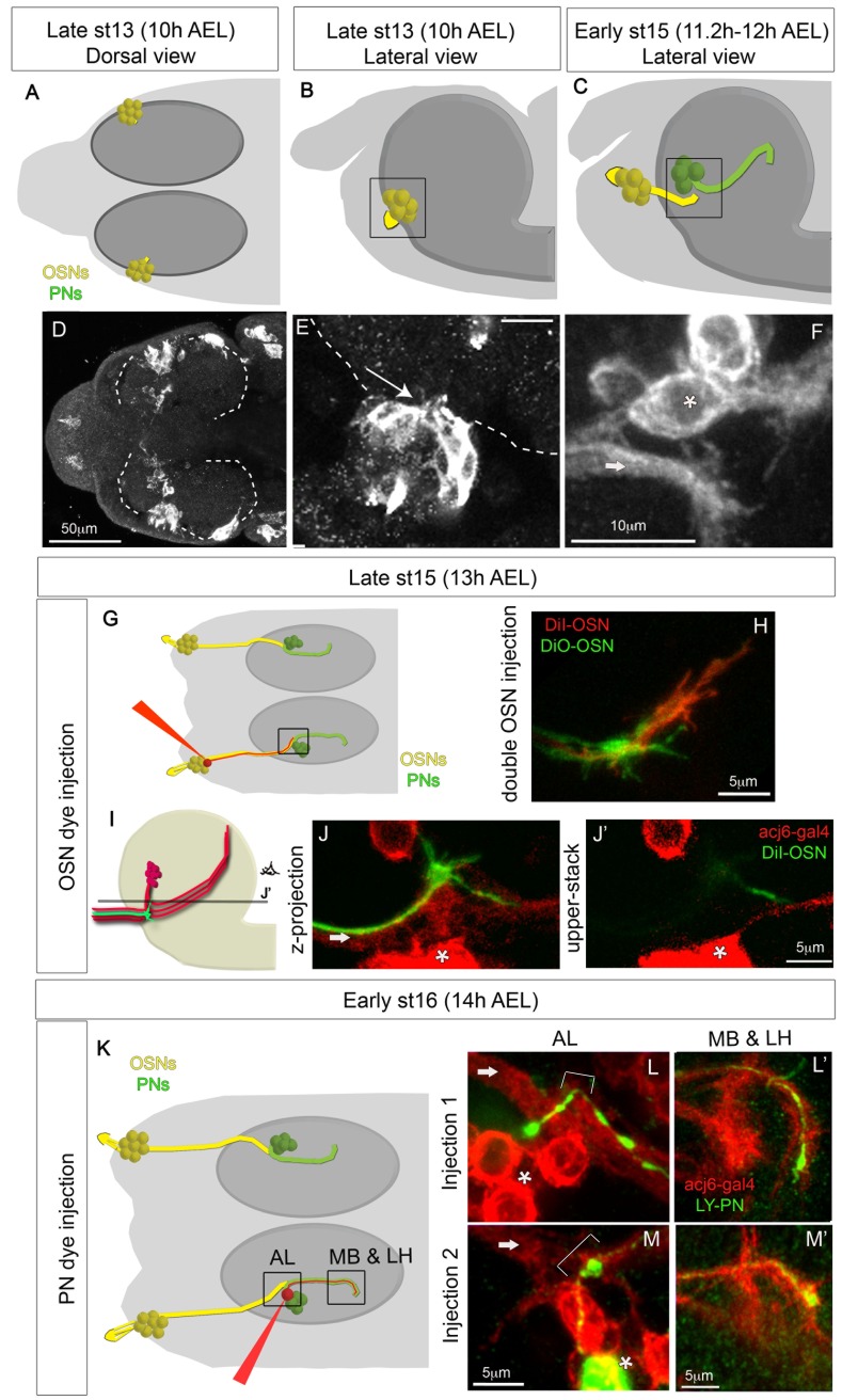
Figure 1. Development of OSNs and PNs between 10 h and 14 h AEL.
(A–F) OSN and PN development as followed with acj6-Gal4:UAS-CD8GFP between 10 h and 12 h AEL, before any contact is made. In the schematics (A–C, G, and K) OSNs are yellow and PNs green. The black square in the schematics indicates the region that is being visualized in the confocal images. In the confocal stacks (E–F, J, L, and M) OSN axons are indicated with an arrow and PN cell bodies with an asterisk. (A–B and D–E) OSNs are born in direct contact with the brain (dashed line) and send short axonal projections into the brain from the very early stages (arrow in E). (F) At early stage 15 (11.2–12 h AEL) OSNs and PNs have not yet contacted each other, although they are within filopodial reach. (G–J) OSN dye injections at 13 h AEL. (G) Schematic showing the injection procedure and marking the region shown in the confocal stacks (H–J) with a black square. (H) Injection of two OSNs with different dyes showing growth cones in both OSNs. (I) Schematic showing the position of OSN axons and PNs. Both components are in red, as in the confocal pictures, showing GFP staining expressed on the acj6-Gal4 pattern. One OSN is green showing filopodia, which represents a single injected OSN as in (J and J′). OSN axons enter the brain in a ventral position respective to PN cell bodies. PN axons run ventrally towards the place of OSN terminals and then turn dorsally towards higher brain centres, making thus a U shape. Different PNs turn their axons at different dorso-ventral depths, and OSN axonal growth cones contact even the most dorsally turning PN axons. The line in (I) marked with the letter J′ represents the confocal z stack shown in (J′). (J) is a confocal z projection of what is represented in (I), while (J′) is a single z stack at the position indicated in (I). From here onwards letters with apostrophe (‘) indicate images taken from the same animal. (K–M) PN dye injections at 14 h AEL. (K) Schematic showing the injection procedure and marking the regions (AL, and MB&LH) shown in the confocal stacks (L–M). (L–M) In each example a single PN was injected (green), and all OSNs and PNs are labelled in red with acj6-Gal4;UAS-CD8GFP. The AL and the MB and LH regions are shown for each injected PN. At 14 h AEL PNs have already extended axons towards higher brain centres, but they have not yet sprouted dendrites at the AL. The axons in the MB and LH are still immature with growth cones. Arrows indicate OSN axons, and brackets indicate the region where the AL is forming and therefore the region where PNs will sprout dendrites.

