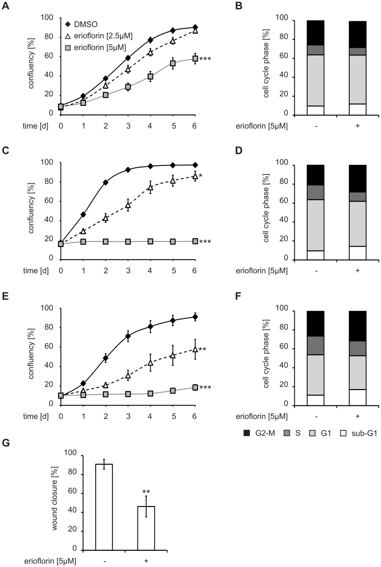
Figure 6. Erioflorin inhibits cell proliferation and alters cell cycle progression.
(A, C, E) MCF7, HeLa and RKO cells were seeded at 10–20% confluency one day prior to the experiment and treated with DMSO (black diamonds) or erioflorin (2.5 (white triangles) and 5 µM (gray squares)). Cell confluency was followed for six days. (B, D, F) MCF7, HeLa and RKO cells were serum deprived for 48 h and treated with erioflorin (5 µM) for 16 h. After propidium iodide staining, distribution of the cells to the different phases of the cell cycle (subG1 (white), G1 (light gray), S (dark gray), G2/M (black)) was determined. (G) RKO colon carcinoma cells were subjected to a scratch wound assay. After administration of the scratch, medium was changed to control or erioflorin (5 µM) containing medium. Wound closure was measured after 24 h and relative wound closure is given as the ratio of the width of the scratch at 24 h and to 0 h. All data are given as means ± SEM (n≥3, *p<0.05, **p<0.01, ***p<0.001).

