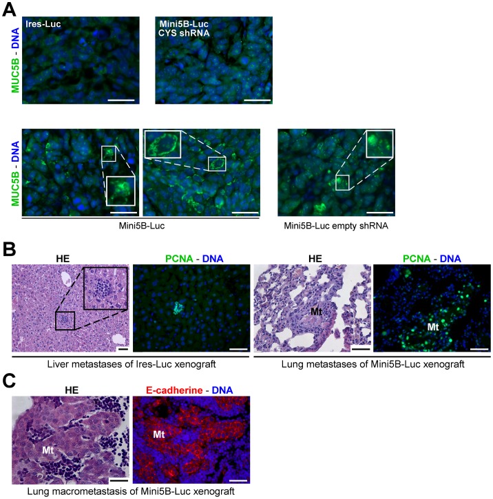
Figure 5. Impact of Mini5B expression on the dissemination of MCF7 cell xenografts in immunodeficient mice.
(A) Immunofluorescence analysis was performed on paraffin-embedded sections of tumors from mice injected with the Ires-Luc clone, the Mini5B-Luc CYS shRNA clone, the Mini5B-Luc clone and the Mini5B-Luc empty shRNA clone and stained with anti-MUC5B antibody. Mini5B was expressed and secreted by tumoral cells in tumors of Mini5B-Luc group and Mini5B-Luc empty shRNA. (B) Serial sections of livers of mice injected with the Ires–Luc clone and stained with HE and anti-PCNA antibody, and serial sections of lungs of mice injected with the Mini5B–Luc clone and stained with HE and anti-PCNA antibody. Metastases in the liver and the lung were visualized. Metastatic cells were PCNA positive. (C) Histological analysis of a macrometastasis in lung of a mouse injected with the Mini5B–Luc clone using HE staining and E-cadherin immunostaining (serial sections). Mt; metastasis. Nuclei were counterstained with Hoechst 33258. Scale bar 50 µm.

