Figure 5.
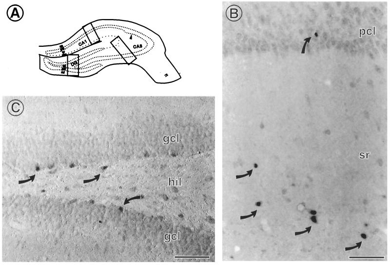
By light microscopy, ERα immunoreactivity (IR) is found in scattered interneurons in the hippocampal formation. (A) Schematic diagram of regions examined by light and electron microscopy. (B) In CA1, a few interneurons with cell nuclear ERα are found primarily in stratum radiatum (sr) and occasionally in the pyramidal cell layer (pcl). (C) Scattered interneurons located within the infragranular regions of the hilus (hil) contain ERα IR associated with their cell nuclei. DG, dentate gyrus; gcl, granule cell layer; CA1, CA3 regions of Ammon's horn; ml, molecular layer; so, stratum oriens; slm, stratum lacunosum moleculare. Scale bars = 40 μm. [Reproduced with permission from ref. 11 (Copyright 2001, Wiley–Liss, Inc., a subsidiary of John Wiley & Sons, Inc)].

