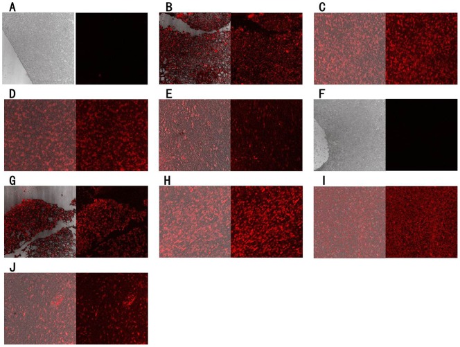
Figure 5. Using the selected aptamers to recognize FFPE normal or glioma tissue sections.
FFPE tissue sections were incubated with cy5-labeled aptamers. A = Normal brain tissue with GBM128; B = Glioblastoma tissue with GBM128; C = Anaplastic oligodendroglima with GBM128; D = Oligoastrocytoma with GBM128; E = Pilocytic astrocytoma with GBM128; F = Normal brain tissue with GBM131; G = Glioblastoma tissue with GBM131; H = Anaplastic oligodendroglima with GBM131; I = Oligoastrocytoma with GBM131; J = Pilocytic astrocytoma with GBM131. The final concentration of Cy5-labeled aptamers was 250 nM.

