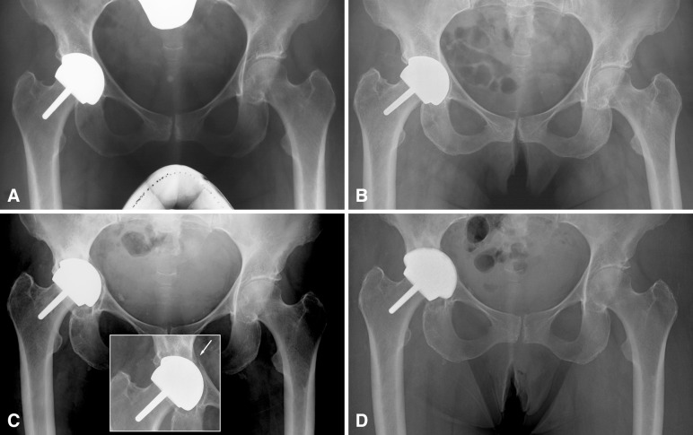
Fig. 3A–D.
(A) Five-month postoperative AP radiograph of a 63-year-old woman who received hip resurfacing for idiopathic osteoarthritis. The 38-mm inside diameter acetabular component was implanted with 56° of abduction and 22° of anteversion, which yielded a contact patch to rim distance of 7.6 mm. The bone-cup interface shows no radiolucencies, suggesting good fixation. (B) Two and a half years after surgery, there is radiolucency visible in DeLee and Charnley Zone III. (C) Nine years after surgery, the radiolucency has expanded around the cup indicating its loosening. The cup has also migrated into the acetabular wall, which showed an unexplained fracture but no complete loss of fixation 6 years after surgery (inset). (D) The reconstruction was maintained as a hip resurfacing and a cup of larger thickness with a titanium backing (Biofoam®; Wright Medical Technology Inc, Arlington, TN, USA) was inserted to articulate with the original resurfacing femoral component.

