Fig. 1.
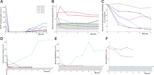
Serial measurements of immune and endocrine parameters. A, Absolute peripheral B lymphocyte numbers over time after rituximab therapy. B lymphocytes were measured as CD19+ cells at flow cytometry (10,000 lymphocyte events counted twice for each measurement; complete depletion was judged as CD19+ < 0.1% of lymphocytes) (Becton Dickinson Instruments, Oxford, UK). The reference range for CD19+ is 90–660 cells/μl. B, The concentration of serum IgM, measured by immunometric nephelometry (BN2; Dade Behring, Surrey, UK), is shown over time after rituximab therapy. The lower limit of the adult female reference range is shown at the top of the gray shaded box (0.72 g/liter), and the lower limit of the adult male reference interval is shown at the top of the hatched box (0.24 g/liter). Three of the four female patients in the study (patients 1, 2, and 5) developed subnormal IgM levels during follow-up. C, The concentration of serum 21-hydroxylase autoantibodies over time is shown as a percentage of the baseline measurement. Measurements were made in duplicate by immunoprecipitation of radiolabeled recombinant in vitro translated 21-hydroxylase protein (RSR Ltd.) (15). For patient 5, sera were diluted 100-fold to measure the concentration. Absolute values of 21-hydroxylase antibody at baseline are shown for each subject in Table 1. D, Serial peak serum cortisol concentrations at diagnosis, at study baseline, and after rituximab treatment. All steroid hormone measurements were made after 38–42 h of withdrawal from regular glucocorticoid and fludrocortisone replacement therapy. For each time point, the highest serum cortisol is recorded, either the baseline sample or after the iv tetracosactide 250 μg (30 or 60 min). The normal response to this stimulus is a peak cortisol concentration greater than 550 nmol/liter (20 μg/dl). Measurements were made using a competitive chemoluminescent immunoassay on a Centaur platform (Siemens, Surrey, UK) with a limit of detection (LD) of 25 nmol/liter (0.91 μg/dl), shown as the shaded horizontal area. To convert serum cortisol from nanomoles per liter to micrograms per deciliter, divide by 27.6. E, Serial recumbent serum aldosterone concentrations at study baseline and after rituximab treatment. The reference range for recumbent serum aldosterone is 100–450 pmol/liter. Measurements were made by solid-phase RIA (DPC Coat-a-Count kit; Diagnostic Products Corp., Surrey, UK), with a LD of 70 pmol/liter, shown as the shaded horizontal area. Several patients' data are superimposed on this limit of detection line. To convert serum aldosterone from picomoles per liter to nanograms per deciliter, divide by 27.7. F, Serial serum DHEAS concentrations at study baseline and after rituximab treatment. Measurements were made by solid-phase competitive chemoluminescence on an Immulite platform (Siemens) with a LD of 0.4 μmol/liter (shown as the shaded horizontal area). The four female patients' data are superimposed on this limit of detection line. The female reference interval is 1.5–10.5 μmol/liter, and the male reference interval is 2.1–12.9 μmol/liter. To convert serum DHEAS from micromoles per liter to micrograms per deciliter, divide by 0.0271.

