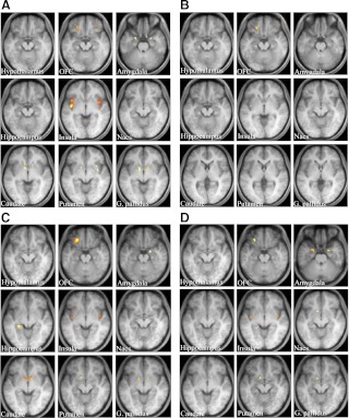
Fig. 1.
Neural response to food-specific stimuli in healthy controls and leptin-off patients. Food-specific activations in ROI in the brains of controls (A and B) and patients (C and D) under fasting (A and C) and postprandial (B and D) conditions. Activation is overlaid onto the group average T1-weighted anatomical axial images (right is right side of the brain). The brighter yellow color represents the higher Z-score. ROI areas are the hypothalamus, orbitofrontal cortex (OFC), amygdala, hippocampus, insula, nucleus accumbens (Nacs), caudate, putamen, and globus pallidus (G. pallidus).

