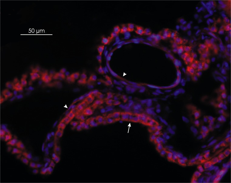
Fig. 1.
Distribution of vascular endothelial growth factor (VEGF-A) in the ovine choroid plexus. The red immunoreactivity of VEGF-A is strongly expressed in cobblestone-shaped epithelial cells (arrow), in comparison with the weaker immunoreactive signal observed in the spindle-shaped endothelial cells (arrowheads). Magnification ×200

