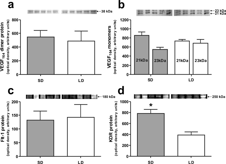
Fig. 5.
Western blot analyses of VEGF-A: VEGF-A164 dimer (a), VEGF-A164 monomers 21 and 23 kDa (b), Flt-1 (c) and KDR (d) in ovine choroid plexuses (CPs) during short days (SD; 8L:16D) and long days (LD; 16L:8D). Upper panels representative blots of CPs resolved by SDS-PAGE and immunoblotted with VEGF-A, Flt-1 and KDR antibodies. VEGF-A was visualized with alkaline phosphatase, whereas Flt-1 and KDR were visualized using the WesternDot™ 625 Western Blot Kit, which requires UV light for visualization. Lower panels mean ± SEM of the densitometric analysis of relative protein levels. *p < 0.05

