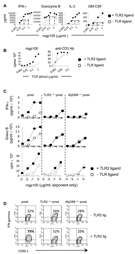
Figure 1. Activating TLR2-MyD88 signals in tumor-reactive CD8 T-cells lowers the activation threshold to a weakly immunogenic tumor-antigen.
Purified pmel, TLR2−/−pmel and MyD88−/− pmel CD8 T-cells were activated with MyD88−/− splenocytes pulsed with varying concentrations of the mgp100 peptide or plate-bound anti-CD3 antibody with or without TLR2 agonist. Four days later cytokine levels determined were by ELISA whereas proliferation was determined by 3H-thymidine uptake. (D) The intracellular level of IFN-γ and granzyme B were determined by flow cytometry 4 days following activation mgp100-pulsed APCs. Shown on the upper right-hand of each plot are the percent of cytokine-positive cells. All data are representative of three or more independent experiments each yielding identical trends.

