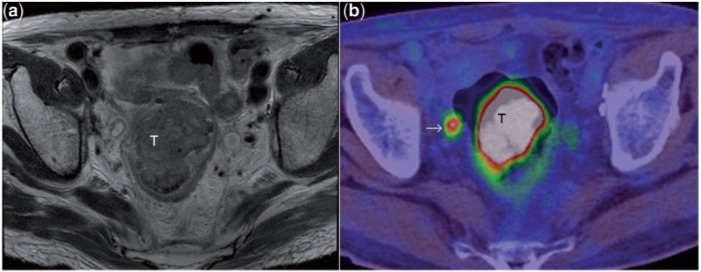
Figure 14.
T2-weighted axial MR image (a) depicts a large rectal mass (T). PET/CT image (b) at a corresponding level shows the intense uptake of fluorodeoxyglucose (FDG) in the tumour. Note also the FDG avid focus (arrow) in the mesorectal fat, consistent with mesorectal lymph node metastasis. In this patient, MRI had shown a few sub-centimetre mesorectal nodes of indeterminate nature. The intense FDG uptake in the node shown confirmed its metastatic nature. Although FDG PET improves the specificity of lymph node characterisation as seen here, the sensitivity is limited[29].

