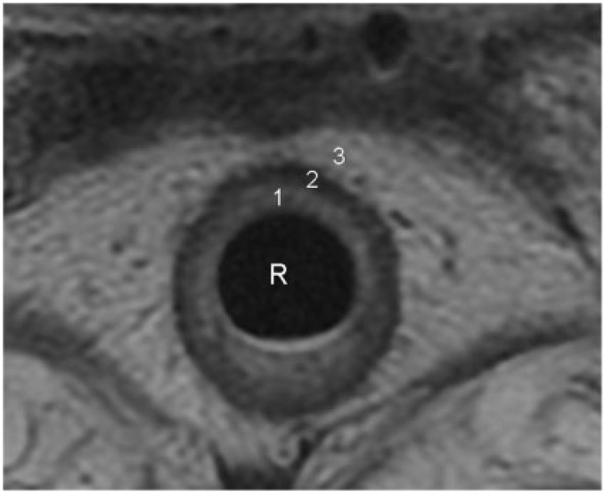
Figure 2.
Concentric layers of normal rectal wall demonstrated on an axial T2-weighted turbo spin-echo MR image. 1, the inner hyperintense layer that represents the mucosa and submucosa; 2, an intermediate hypointense layer that represents the muscularis propria; 3, an outer hyperintense layer that represents the perirectal fat. Further differentiation between mucosa and submucosa is occasionally possible when the submucosa is identified as a markedly hyperintense layer sandwiched between the mucosa and muscularis propria. R, rectal lumen.

