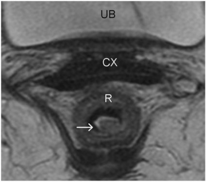
Figure 4.
Rectal carcinoma in situ. High-resolution axial turbo spin-echo T2-weighted MR image shows a polypoid rectal lesion (arrow). The intact muscularis propria at the level of the stalk of the polypoid tumour confidently excludes T2 disease. Involvement of submucosa cannot be excluded and hence was staged as T1 preoperatively. However, at surgical pathology this lesion was staged as carcinoma in situ. The differentiation between carcinoma in situ and a T1 tumour is difficult, especially when the lesions are sessile and when the mucosa and submucosa are not identified separately (a common finding). R, rectum; CX, cervix; UB, urinary bladder.

