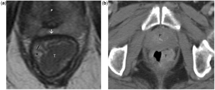
Figure 5.
Stage T1 rectal tumour. (a) Axial T2-weighted MR image shows a large rectal tumour of intermediate signal intensity. The rectal mucosa (parallel lines), submucosa (asterisk) and muscularis propria (arrow) are distinctly visualised in the high-resolution MR image. The tumour involves the mucosa and submucosa while the muscularis propria is preserved, consistent with T1 disease. (b) Post-contrast axial CT image at a level corresponding to that in (a) demonstrates the tumour but gives limited information on the depth of invasion. The limited soft-tissue resolution (compared to MRI) of CT precludes confident T-staging of the rectal tumour. T, tumour; P, prostate.

