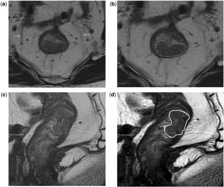
Figure 6.
Stage T2 rectal tumour. (a, b) High-resolution T2-weighted axial MR images show the rectal tumour invading the muscularis propria posteriorly between the 4- and 8-o’clock positions. A thin, relatively hypointense stripe representing the muscularis propria is interposed between the mesorectal fat and as demonstrated in high- resolution T2-weighted sagittal MR images (c, d). (b) The tumour invades the muscularis propria but spares the mesorectum; hence it is staged as T2 disease. T, tumour. Asterisks indicate mesorectum; arrows indicate mesorectal fascia; tumour outlined in white in (b) and (d).

