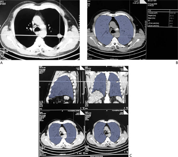
Figure 1.

Quantitative Ct Volume Estimations. (A): Chest CT scan of a patient with a tumor in the left upper lobe. (B): and (C): Quantitative analysis of functional lung parenchyma of both lungs, using the dual threshold of -500 to -910 HU. Areas in blue correspond to voxels within these attenuation limits. Total functional lung volume of both lungs is estimated to be 3932.99 mL.
