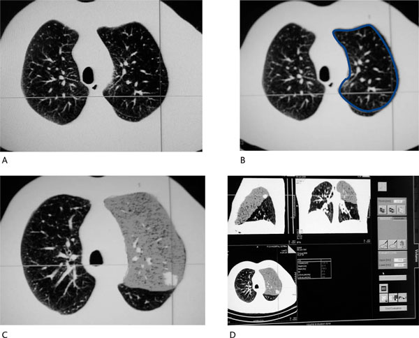
Figure 2.

Volumetric Analysis of the Resected Lobe (Same Patient as in Figure 1). (A): Fissure identification between left upper and lower lobe. (B): Delineation of the region of interest (limits of the lobe to be resected) in all transaxial images. (C): and (D): Volumetric analysis of the left upper lobe. Regional functional lung volume is estimated to be 1173.14 mL.
