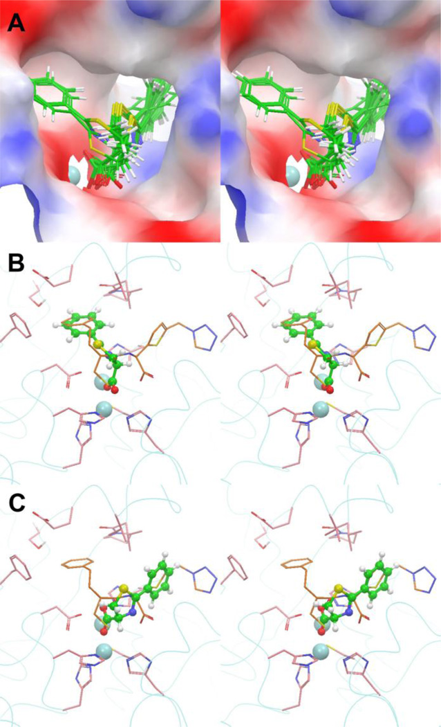
Figure 3.
(A) Stereoview of the 20 lowest-energy docking structures of compound 4 in the active site of the crystal structure of IMP-1:MCI complex, with the protein shown as an electrostatic potential surface and Zn2+ as a light blue sphere; (B) The lowest-energy docking structure of conformation A of 4 (the ball and stick model in green) in the active site, superimposed with the crystal structure of MCI (in orange); (C) The lowest-energy docking structure of conformation B of 4 superimposed with the structure of MCI.

