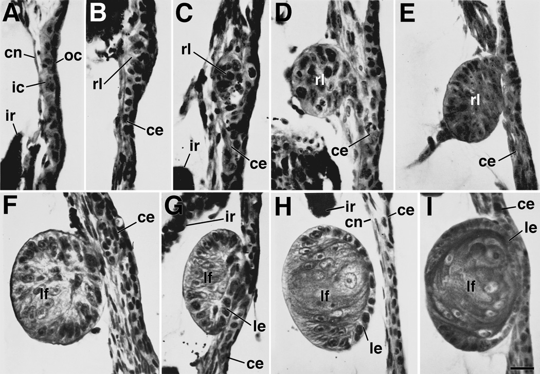
Figure 1.
Cornea-lens transdifferentiation in X. laevis. Sections show different stages of lens regeneration following removal of the lens (stages follow the convention of Freeman,1963). A. Stage 1. During this stage cells of the inner corneal epithelium assume a cuboidal shape within 24 hours following lens removal. B. Stage 2. During this stage, cells begin to assume a thickened placodal arrangement. Nuclei of these cells are found to contain one nucleolus (characteristic of lens epithelial cells), rather than two (characteristic of cornea epithelial cells). C. Early stage 3. D. Middle stage 3. E. Late stage 3. During stage 3, a loosely organized aggregate of cells begins to separate from the corneal epithelium. These cells begin to organize with distinct apical-basal polarity to form a lens vesicle. F. Early stage 4. G. Middle stage 4. H. Late stage 4. During stage 4 a definitive lens vesicle is present. Elongated primary lens fiber cells have formed on the side closest to the vitreous chamber. Typically the lens vesicle is separated from the overlying corneal epithelium at this stage. I. Early stage 5. During this stage, secondary lens fiber cells are being added from the lens epithelium at the equatorial zone. Nuclei of the primary fiber cells begin to disappear. This stage is followed by further addition of secondary fiber cells and growth of the lens. ce, corneal epithelium; cn, corneal endothelium; ic, inner layer of the corneal epithelium; ir, iris; oc, outer layer of the corneal epithelium; le, lens epithelium; lf, lens fibers; rl, regenerating lens vesicle. Refer to text for further details. Figure from Henry (2003, after Freeman, 1963). Scale bar equals 25µm.

