
Figure 4.
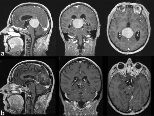
(a) Gadolinium-enhanced T1-weighted MR images obtained in a 55-year-old man (case 2), revealing a large meningioma of pineal region beneath the tentorium and extending to the midbrain. (b) Postoperative MRI after resection via an occipital transtentorial approach