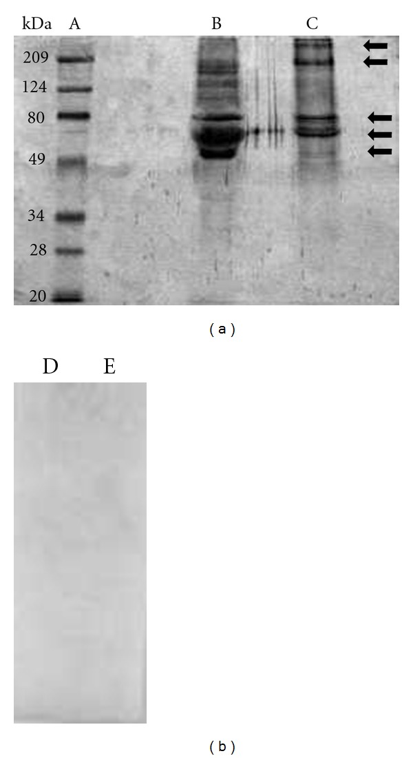
Figure 2.

Western blot analysis of sodium dodecyl-polyacrylamide gel electrophoresis-separated ESEAs: Au and RG1 T. cruzi isolates. (A) standard, (B) RG1 isolate, and (C) AU isolate. The dot was probed with a pool of sera from patients with Chagas disease (B and C) and with a pool of control-negative sera RG1 and Au isolate, (D and E), respectively.
