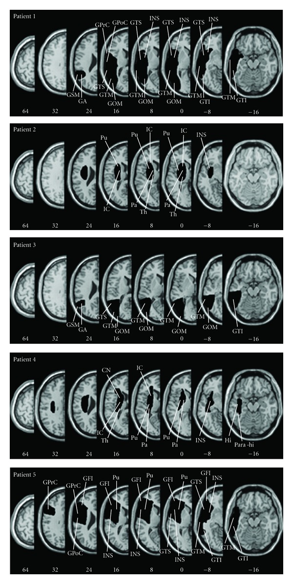
Figure 1.

Lesion anatomy. For each patient, all lesions were mapped using the free MRIcro software and were drawn manually on slices of the high-resolution 3D T1-weighted template MRI scan. This template is oriented to match the Talairach space. Lesions were mapped onto the horizontal slices that correspond to Z-coordinates −16, −8, 0, 8, 16, 24, 32, 64 in the Talairach space by using the identical or the closest matching horizontal slices of each individual. Following radiological convention, the right cerebral hemisphere is displayed on the left side. Abbreviations: CN, caudate nucleus; GFI, gyrus frontalis inferior; GOM, gyrus occipitalis medius; GPrC, gyrus precentralis; GPoC, gyrus postcentralis; GTM, Gyrus temporalis medius; GTI, gyrus temporalis inferior, GTS, gyrus temporalis superior; Hi, hippocampus; IC, internal capsule; INS, insula; Pa, pallidum; Para-hi, parahippocampus; Pu, putamen; Th, thalamus; GSM, gyrus supra-marginalis; GA, gyrus angular.
