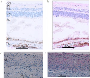
Fig. 3.
CYP4V2 expression in normal human retina and cornea. Immunohistochemical staining with the anti-CYP4V2 IgG showed strong positive staining of RPE cells in retinal tissue and weak staining of ganglion cells and internal/external nuclear layers in the retina and corneal epithelial cells. a, retina treated with preimmune IgG; b, retina treated with anti-CYP4V2 IgG; c, cornea treated with preimmune IgG; d, cornea treated with anti-CYP4V2 IgG. GCL, ganglion cell layer; IPL, inner plexiform layer; INL, inner nuclear layer; ONL, outer nuclear layer; PhL, photoreceptor layer; Ch, choroid; AE, anterior epithelium; SP, substantia propria.

