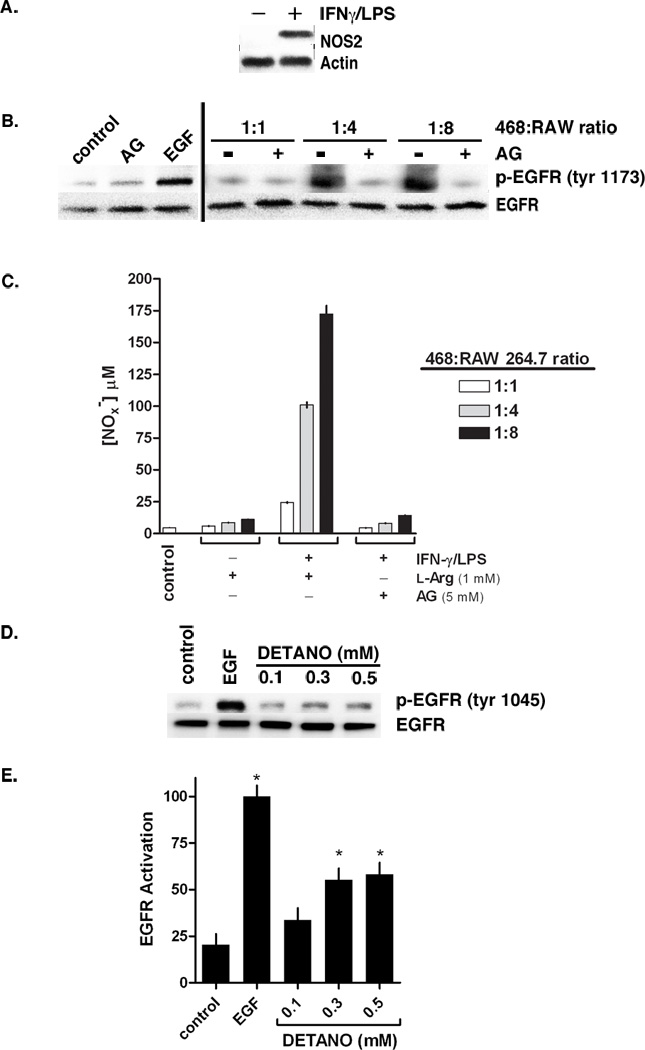
Figure 1. NOS2 activity and NO-donor increases EGFR tyrosine phosphorylation.

(A) Relative NOS2 expression in RAW 264.7 murine macrophages pretreated with or without IFN-γ/LPS. (B) Western blot showing EGFR tyrosine 1173 phosphorylation in MDA-MB-468 cells co-cultured with IFN-γ/LPS activated RAW cells in increasing ratios and in media containing or lacking the NOS2 inhibitor aminoguanidine (AG). Co-cultured samples are compared to monocultured MDA-MB-468 cells treated with AG or EGF. (C) Total nitrate and nitrite (NOx−) concentrations in conditioned media from co-culture experiments as an aggregate indicator of NOS2 activity. (D) Western blot of EGFR tyrosine 1045 phosphorylation in serum-starved MDA-MB-468 cells treated with either EGF (10 ng/ml) or DETANO for 24 hours. (E) Densitometric analyses of EGFR tyrosine 1173 phosphorylation indicating that EGF and 0.3 and 0.5 mM DETANO resulted in significant increases in EGFR activation compared to untreated controls (*P < 0.05).
