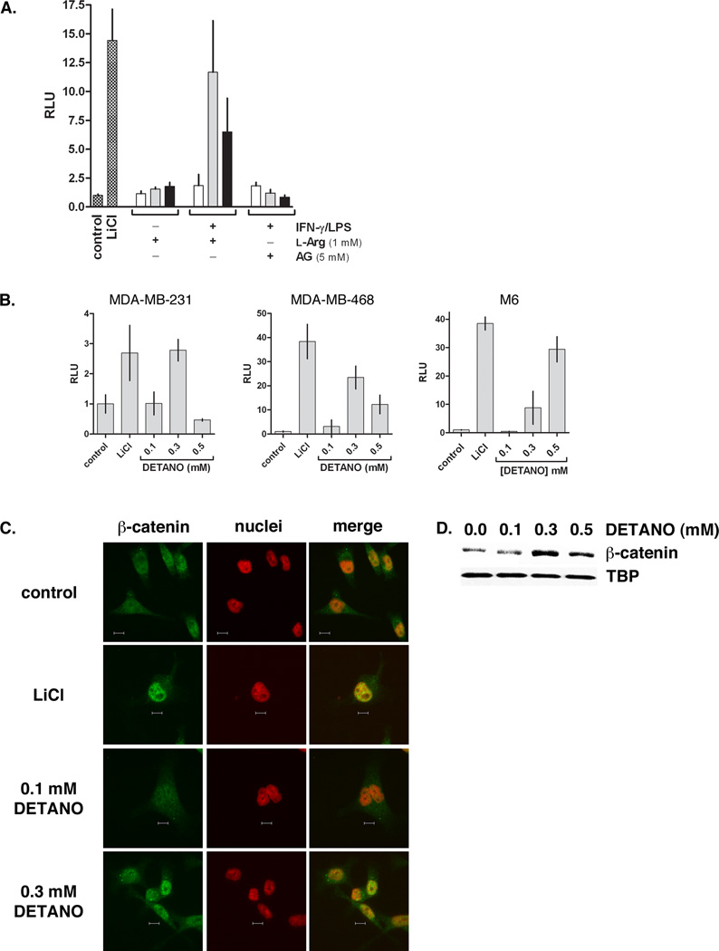
Figure 4. NO activates the β-catenin signaling pathway in basal-like breast cancer cells.
(A) TCF-luciferase reporter activity of MDA-MB-468 cells alone or co-cultured with RAW 264.7 cells under the conditions shown. Data are normalized to untreated controls and represent the fold-increase of mean relative luminescence units (RLU) (± sd). (B) TCF-luciferase reporter activity from breast cancer cell lines (MDA-MB-468, MDA-MB-231 and M6) exposed to LiCl or DETANO for 20 hours. Data represent mean fold-increase RLU (± sd) compared to untreated controls. (C) Representative immunofluorescent micrographs of MDA-MB-231 β-catenin cellular localization in response to LiCl or DETANO exposure. The yellow pixels in merged images represent nuclear β-catenin localization and the scale bars represent 10 µm. (D) Representative western blot of MDA-MB-468 nuclear fractions showing an increase in nuclear β-catenin in response to DETANO compared to tata-binding protein (TBP) nuclear content.

