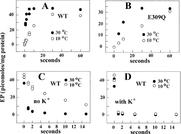
FIGURE 6.
Time course of EP formation through utilization of [32P]Pi by WT SERCA (A) and E309Q mutant (B) and decay (C and D) of [32P]phosphoenzyme after a chase with nonradioactive Pi. Microsomes derived from COS1 cells expressing SERCA or E309Q mutant were incubated with 50 mm MES, pH 6.0, 20% Me2SO4, 10 mm MgCl2, and 2 mm EGTA. The reaction was started by the addition of 50 μm [32P]Pi, quenched at different times, and processed for determination of radioactive protein by electrophoretic analysis and detection of radioactivity (see “Experimental Procedures”). For determination of decay, the protein was incubated with 50 μm [32P]Pi as described above at 10 °C for 2 min, and then a 20-fold dilution was obtained with a medium containing 50 mm MES, pH 6.0, 10 mm MgCl2, 2 mm EGTA, and 1 mm nonradioactive Pi at 10 or 30 °C. Serial samples were taken at sequential times for acid quenching and determination of residual radioactive protein by electrophoretic analysis and detection of radioactivity. The experimental points are averages of values obtained in three to four different experiments.

