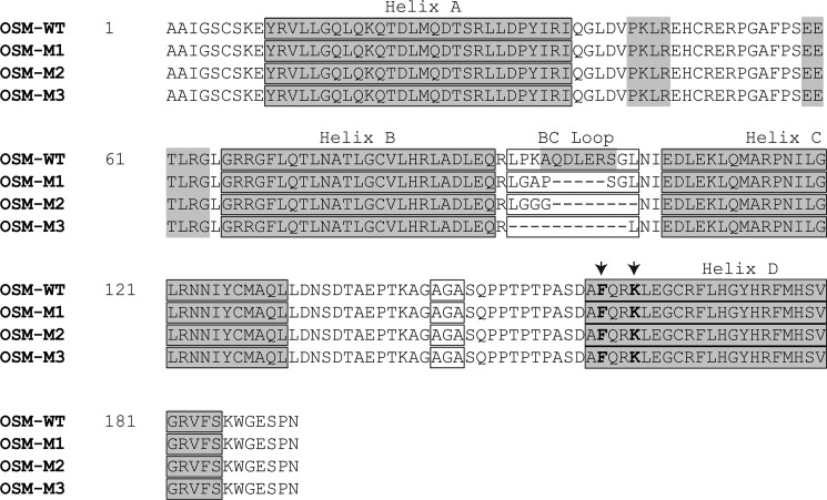
FIGURE 2.
Amino acid sequences of wild-type OSM and the mutant variants of OSM with truncated BC loops. Shown in gray are the α-helices present in the secondary structure of OSM as identified in the crystal structure (PDB 1EVS). Each of the helices A, B, C, and D are identified along with the BC loop region. Also highlighted in an open box is the mutated thrombin cleavage site AGA, and shown in bold letters indicated by arrows is the active FXXK site on the wild-type and mutant OSMs required by the molecules to bind LIFR and OSMR.

