FIGURE 10.
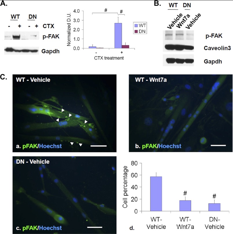
Sulfs promote FAK activation and clustering. A, TA muscles of WT and SulfSK-DN mice were collected at day 3 post-injury for Western blot assays of p-FAK. Gapdh was the loading control. Images represent one of four independent experiments. B, WT and SulfSK-DN myoblasts in cultures were treated with vehicle or Wnt7a (100 ng/ml) for 48 h before Western blot assays of p-FAK and caveolin3. Gapdh was the loading control. Images represent one of three independent experiments. C, shown is immunocytochemistry to assay subcellular localization of p-FAK (green) in WT myoblast cells that were treated with vehicle or Wnt7a for 48 h and in SulfSK-DN myoblasts in differentiation medium. Nuclei were labeled with Hoechst dye (blue). Arrowheads point to punctuated p-FAK clustering. Percentages of cells with punctuated p-FAK immunolabeling are shown in d. Data are the mean and S.D. of four independent experiments. #, p < 0.01. Scale bars, 100 μm.

