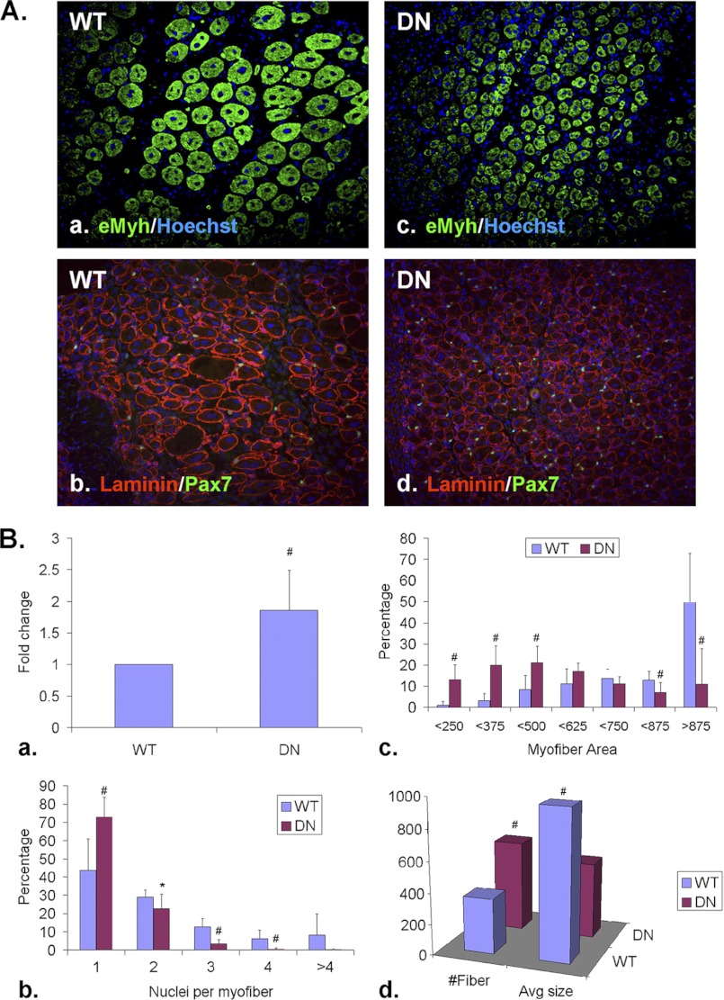
FIGURE 2.
Sulfs promote SC differentiation and myoblast fusion during skeletal muscle regeneration. TA muscles of WT and SulfSK-DN mice were harvested at day 5 post-injury. A, Pax7+ SCs and eMyh+ newly formed myofibers were identified by immunohistochemistry using mid-belly cross-sections. The basement membrane of myofibers was labeled with a laminin antibody. Nuclei were labeled by Hoechst dye. Scale bar = 100 μm. B, shown is quantification of relative abundance of Pax7+ SCs (a), relative abundance of new myofibers with increasing number of myonuclei (b) and sizes (c), and average number and size of new myofibers within a regenerating area of 1 mm2 (d) of WT and SulfSK-DN muscles. Data presented are the mean and S.D. of more than 1000 new myofibers from 3 independent experiments. #, p < 0.01; **, p < 0.05; *, p < 0.1.

