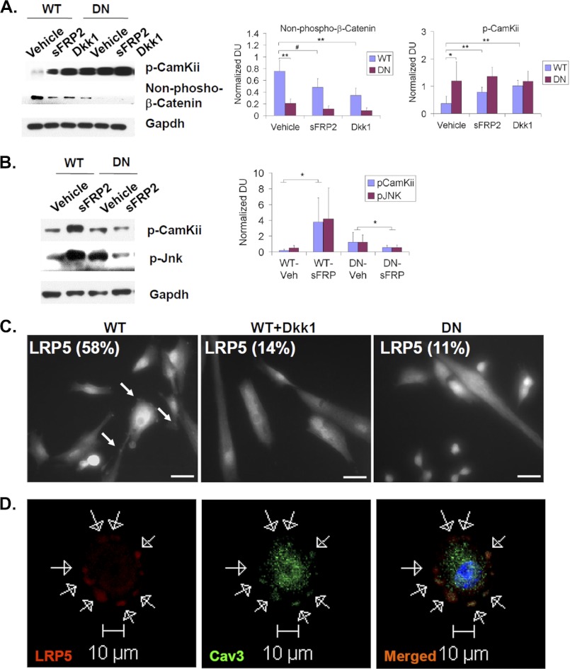
FIGURE 7.
Sulfs indirectly repress noncanonical Wnt signaling but directly promote opposing canonical Wnt signaling. A and B, primary myoblast cultures (in A) or regenerating TA muscles (in B) of WT and SulfSK-DN mice were treated with Wnt inhibitors (2 μg/ml sFRP2 or 0.5 μg/ml Dkk1). Downstream activators of canonical and noncanonical Wnt signaling, stabilized β-catenin and p-CamKii/p-Jnk, respectively, were assayed by Western blot. Gapdh was the loading control. Quantification was performed by normalizing signals of p-CamKii and stabilized β-catenin to Gapdh by densitometry. Images represent one of four independent experiments. C, primary WT myoblast cultures of WT and SulfSK-DN mice in growth medium were immunolabeled using an antibody against LRP5. WT myoblast cells exhibited punctuated LRP5 labeling (pointed by white arrows), whereas LRP5 labeling appeared diffusive in DKK1-treated WT myoblast cultures and in SulfSK-DN myoblast cultures. The percentage of cells that exhibited punctuated LRP5 staining was shown in images. Images shown were representative of three independent experiments. Scale bars, 50 μm. D, colocalization of LRP5 and caveolin3, a lipid raft marker, in WT myoblasts by confocal microscopy is shown. Arrows pointed to punctuated immunolabeling of LRP5 and caveolin3 around the cell surface. #, p < 0.01; **, p < 0.05; *, p < 0.1. DU, density unit.

