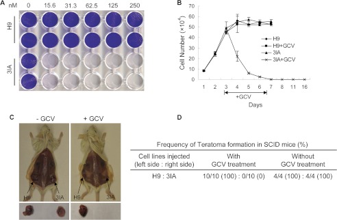
FIGURE 2.
GCV eliminates TK-hESCs (3IA) in vitro and abolishes the teratoma formation of TK-hESCs in vivo. A, TK-hESCs are hypersensitive to GCV. H9 and 3IA TK-hESCs were treated with increasing concentrations of GCV. Three days after GCV treatment, cells were fixed and stained with crystal violet. The genotypes are indicated on the left, and the concentrations of GCV (nm) are indicated on the top. B, TK-hESCs were rapidly eliminated by GCV in culture. H9 hESCs and TK-hESCs were plated, mock-treated, or treated with 0.25 μm GCV for 4 days. GCV was removed from the medium, and the cells were cultured for additional 9 days. Cell numbers were counted at the indicated time points. C, GCV selectively abolishes the teratoma formation of TK-hESCs in SCID mice. SCID mice were subcutaneously injected with H9 hESCs and TK-hESCs around the left and right hind legs, respectively. One day after implantation, mice were intraperitoneally injected with GCV of various dosages for one or 2 weeks. Six-to-8 weeks after implantation, SCID mice were euthanized, and teratomas were examined. Representative images are shown. D, the summary of the frequency of teratoma formation of hESCs and TK-hESCs in SCID mice with or without GCV treatment.

