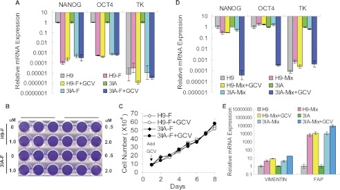
FIGURE 3.
Fibroblasts (indicated by F) differentiated from TK-hESCs (3IA) are insensitive to GCV. A, the relative mRNA levels of NANOG, OCT4 and TK in fibroblasts derived from the teratomas formed by H9 and TK-hESCs. Fibroblasts were treated with or without 1.0 μm GCV for 4 days. The mRNA levels in fibroblasts are compared with those in hESCs. Mean value are presented with S.D. (n = 3). B, fibroblasts derived from H9 and TK-hESCs were treated with increasing concentrations of GCV for 4 days and subsequently stained with crystal violet. C, the proliferation curve of fibroblasts derived from H9 hESCs and TK-hESCs with or without 1.0 μm GCV treatment. Cells were treated with GCV, and duplicate wells were counted at the indicated time points. D and E, GCV treatment selectively eliminates undifferentiated TK-hESCs when spiked into fibroblast culture. hESCs were plated onto a six-well plate with 1 × 10⁵ cells per well together with 2 × 10⁵ fibroblasts and treated with 1.0 μm GCV for 4 days. The mRNA levels of the NANOG, OCT4, TK, as well as vimentin and fibroblast activation protein (FAP) by the remaining cells were analyzed by qPCR and compared with those in hESCs. Mean value are presented with S.D. (n = 3).

