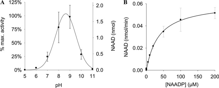
FIGURE 3.
Characterization of NAADP-degrading activity in HeLa cells. A, pH dependence of NAADP-degrading activity. Membrane protein from HeLa cells (1 mg/ml) was incubated with 20 μm NAADP at 37 °C for 5 min at different pH values. Reaction products were analyzed by HPLC. Buffers used were as follows: pH 5–6, MES; pH 7–9, TEA buffer; and pH 10–11, DEA buffer. Data shown are means ± S.D. (n = 2–4; p < 0.001). B, kinetic characterization of NAADP-degrading activity. Membrane protein from HeLa cells (0.5 mg/ml) was incubated with increasing concentrations of NAADP in TEA buffer at pH 9. Reaction products were analyzed by HPLC. Data shown are means ± S.D. (n = 1–6).

