
FIGURE 6.
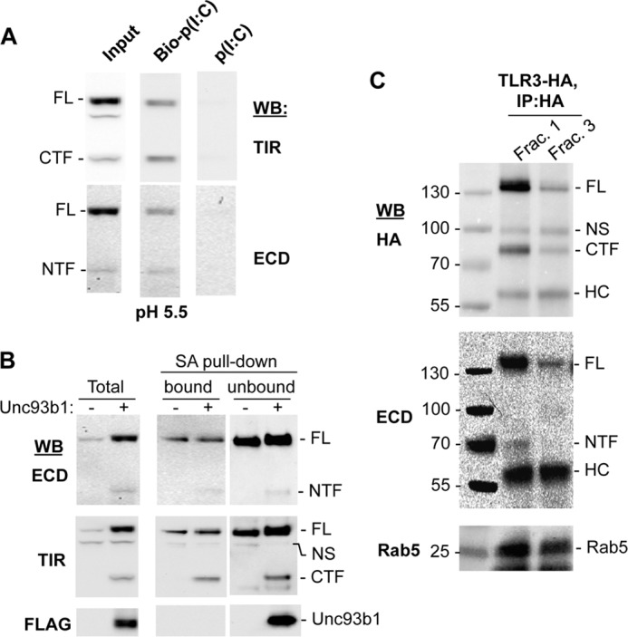
The proteolyzed TLR3 fragments are on the surface, in endosomes, and can bind poly(I:C). A, the TLR3 NTF and CTF retain the ability to bind poly(I:C). HEK293T cells transfected to express TLR3 and Unc93b1 were lysed in acidic buffer, pH 5.5. Poly(I:C) or biotinylated poly(I:C) was incubated with cell lysates and precipitated with streptavidin-agarose beads. The bound materials were detected using Western blots (WB) probed with antibodies to ECD or TIR of TLR3. B, fragments of TLR3 can be detected on the cell surface. HEK293T cells transfected to express TLR3 with or without coexpressed Unc93b1 were labeled with sulfo-NHS-Biotin, which is impermeable to cell membranes. The streptavidin-agarose beads precipitated (bound) and the remaining supernatant not bound to the beads (unbound) as well as the total cell lysate (Total) were all analyzed for TLR3 and the cleaved fragments by Western blots. C, uncleaved and cleaved TLR3 are enriched in endosomes. HEK293T cells expressing TLR3-HA were homogenized, and organelles were fractionated using an iodoxanol gradient. Fractions that are positive for the endosome marker Rab5 in Western blots (Frac. 1 and Frac. 3) were immunoprecipitated with HA antibody, and TLR3 was detected by Western blots. NS, nonspecific band; HC, heavy chain.