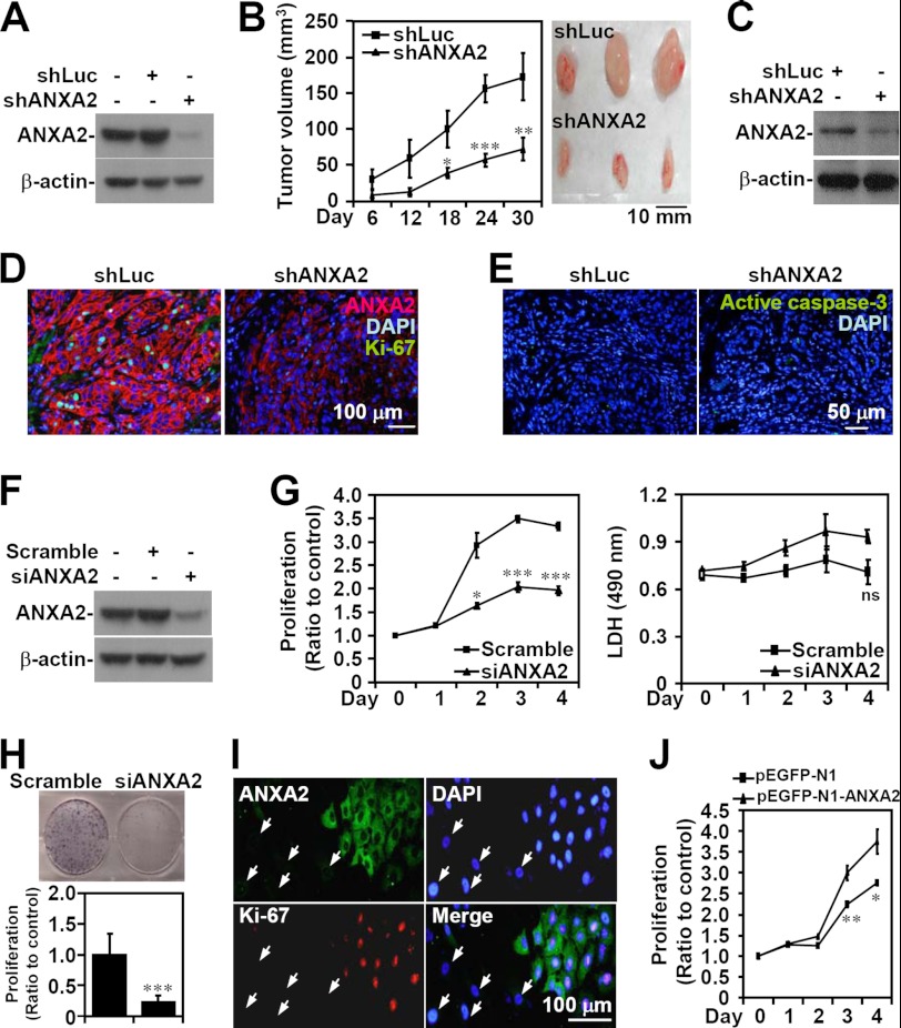
FIGURE 2.
ANXA2 controls cell growth, but not cell survival, in NSCLC. ANXA2 (ANXA2) was silenced in human NSCLC A549 cells by lentivirus-based short hairpin transfection (shANXA2). An shRNA targeting luciferase (shLuc) was used as a negative control. Western blot analysis was used to detect the expression of ANXA2 in transfected A549 cells (A) and tumors from BALB/c nude mice 30 days postinoculation with shLuc- or shANXA2-transfected cells (C). B, tumor growth was analyzed in xenografts with a subcutaneous injection of A549 cells transfected with shLuc or shANXA2 for the indicated time. Tumor volume was calculated (n = 5/group), and the representative morphology of the tumors was photographed (n = 3/group). D and E, sections of subcutaneous tumor were stained for ANXA2 (red), Ki-67 (green), active caspase-3 (green), and nuclei using DAPI (blue). F, small interfering RNA (siANXA2) was also transfected to silence ANXA2 in A549 cells. A non-targeting siRNA (scramble) was used as a negative control. Western blot analysis was used to detect the expression of ANXA2. G, after transfection for the indicated time, cell proliferation and cytotoxicity were determined by WST-8 and lactate dehydrogenase assays, respectively. The data are the means ± S.D. (error bars) of triplicate cultures. *, p < 0.05; ***, p < 0.001 compared with the control. ns, not significant. H, colony formation was used to measure the growth of siANXA2-transfected A549 cells 10 days post-transfection. The data are the means ± S.D. of triplicate cultures. ***, p < 0.001 compared with scramble. I, immunostaining followed by fluorescence microscopy was used to determine the expression of ANXA2 (green) and Ki-67 (red) in siANXA2-transfected A549 cells. The arrows indicate successfully transfected cells. DAPI (blue) was used for nuclear staining. One representative image of three individual experiments is shown. J, ANXA2 was overexpressed in A549 cells by pEGFP-N1-ANXA2 transfection. A WST-8 assay was used to measure the growth of transfected cells. pEGFP-N1 was used as the negative control. For Western blot analysis, β-actin was used as an internal control. One representative data set of three individual experiments is shown.

