FIGURE 2.
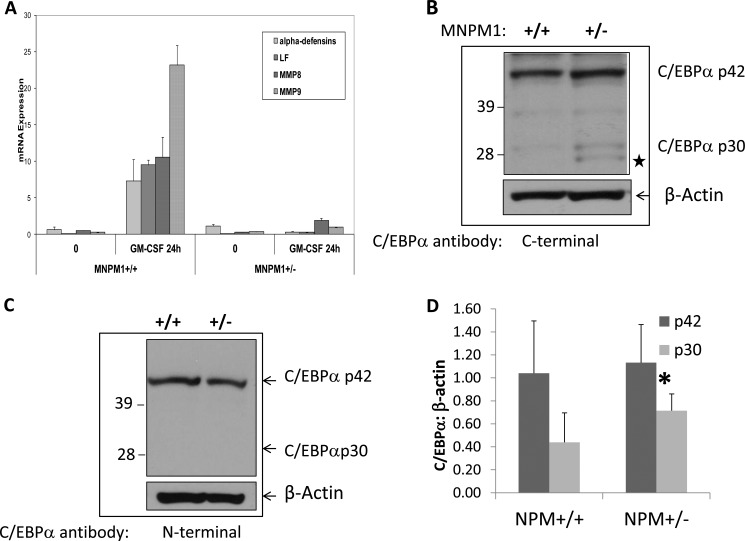
A, neutrophil-specific gene expression is impaired in NPM1+/− myeloid cells. Expression levels of four neutrophil-specific genes, α-defensins, LF, neutrophil collagenase (MMP8), and neutrophil gelatinase (MMP9) were measured using real time PCR analysis using cDNA derived from total RNA from uninduced (0 h) and 48-h GM-CSF-induced MNPM1+/+ and MNPM1+/− cell lines. Expression levels were normalized to that of β-actin. The expression of each gene was measured in triplicate, and the S.E. values are indicated. These data are representative of a total of three independent experiments. B, NPM1 haploinsufficient cells express increased levels of the C/EBPα p30 isoform. Expression levels of C/EBPα were measured by Western blot analysis in uninduced MNPM1+/+ and MNPM1+/− cell lines and probed sequentially with a C-terminal specific C/EBPα antibody and β-actin antibody. This experiment has been repeated on three independent occasions. ★ represents a shorter C/EBPα “isoform” (see supplemental Fig. S1 for details) C, Western blot analysis of cell lysates prepared from MNPM1+/+ and MNPM1+/− cells was performed. The blot was probed sequentially using an antibody specific for C/EBPαp42 recognizing the N terminus of the C/EBPα protein and β-actin antibody as a loading control. Note that no C/EBPαp30 was observed in this blot. D, quantitative analysis of the relative expression levels of C/EBPαp30 and C/EBPαp42 from three independent experiments compared with β-actin expression using Image J software in MNPM1+/+ versus MNPM1+/− cells. No change in expression of p42 was observed. p30 levels increased 1.63-fold in MNPM1+/− cells (*, p = 0.05).

