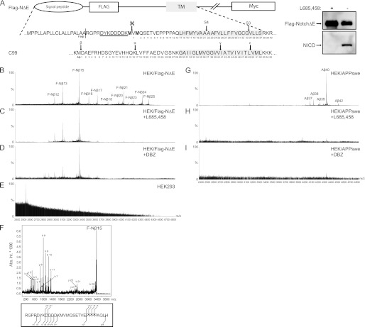
FIGURE 1.
Detection of secreted FLAG-Nβ and Aβ peptides. A, schematic representation of the human Notch1-ΔE protein containing an N-terminal FLAG epitope (FLAG-NΔE) to facilitate detection of the secreted FLAG-Nβ peptides. The two methionine residues depicted in bold are incorporated to prevent S2 cleavage and thus removal of the FLAG epitope. The α-FLAG antibody M2 recognition site is underlined. The part of C99 that is cleaved by β- and γ-secretase is aligned with FLAG-NΔE in respect to transmembrane domains. Arrows show the S2, S3, S4, β, α, γ, and ϵ cleavage sites, and numbering is shown for F-Nβ and Aβ. There is protein expression of HEK293 cells stably expressing human FLAG Notch1-ΔE and the generation of NICD, using a α-FLAG or the Val-1744 antibody. The NICD formation was abolished in the presence of the GSI L-685,458. B, MALDI-TOF MS spectrum of F-Nβ using conditioned medium from HEK/FLAG-NΔE cells that was immunoprecipitated with α-FLAG M2-agarose. F-Nβ numbering, using the nomenclature set by Okochi et al. (40) of the individual peaks are indicated. C and D, MALDI-TOF MS spectrum of F-Nβ using conditioned medium from HEK/FLAG-NΔE cells treated with the GSIs L-685,458 or DBZ, respectively. Spectra analysis reveals that only F-Nβ16–25 is inhibited by GSIs. E, MALDI-TOF MS spectrum of F-Nβ using conditioned medium from HEK293 cells. F, all F-Nβ peaks were identified by MS/MS, and a representative spectrum of F-Nβ15 is shown. G, MALDI-TOF MS spectrum of Aβ using 4G8 immunoprecipitated conditioned medium from HEK/APPswe cells. H and I, MALDI-TOF MS spectrum of Aβ using conditioned medium from HEK/APPswe cells treated with the GSIs L-685,458 or DBZ, respectively.

