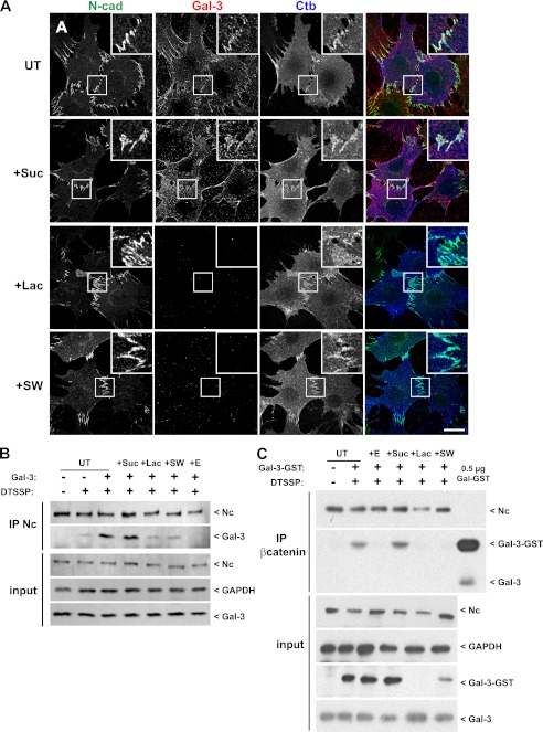
FIGURE 1.
N-cadherin, Gal-3, and Ctb localize to cell-cell junctions. A, Mgat5+/+ cells were untreated (UT) or treated for 48 h with 20 mm sucrose (+Suc), 20 mm lactose (+Lac), or 1 mm swainsonine (+SW) before incubation for 20 min at 4 °C with FITC-coupled Ctb (blue) and cyanine-3-coupled Gal-3 (red). Cells were fixed and stained for N-cadherin (N-cad) (green). Insets show accumulation of Gal-3 and Ctb at N-cadherin-positive cell-cell junctions. Gal-3 binding to the cells is reduced by lactose and swainsonine. Bar: 20 μm. B, Mgat5+/+ cells were untreated or treated for 48 h with 20 mm sucrose, 20 mm lactose, or 1 mm swainsonine and then incubated or not with 1.5 mm EGTA (+E) for 1 h before incubation for 20 min at 4 °C with Gal-3. Cells were then incubated with 0.1 mg/ml DTSSP for 1 h at 4 °C. After quenching and cell lysis, cell extracts were submitted to N-cadherin immunoprecipitation (IP Nc). Pulldowns and total cell extracts (input) were analyzed by Western blot for N-cadherin (Nc) and Gal-3, and inputs were blotted for GAPDH as a loading control. C, Mgat5+/+ cells were treated as described in B except that cells were incubated with GST-Gal-3 and immunoprecipitated with antibodies to β-catenin. 0.5 μg of purified Gal-3-GST were also loaded for Western blot analysis.

