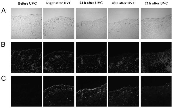
Figure 4.
Immunohistochemical analyses of DNA lesions in a representative mouse skin abrasions after UVC exposure. Biopsies were taken before, immediately after, 24 hours, 48 hours, and 72 hours after UVC irradiation. (A) Micrographs of the morphologies of skin abrasion. (B) Micrographs of 4′,6-diamidino-2-phenylindole counterstaining of cell nuclei. (C) Immunofluorescence micrographs of CPDs in skin cell nuclei. Color figure available online as Supplemental Digital Content 3, http://links.lww.com/TA/A183.

